Confocal microscopes

More Information

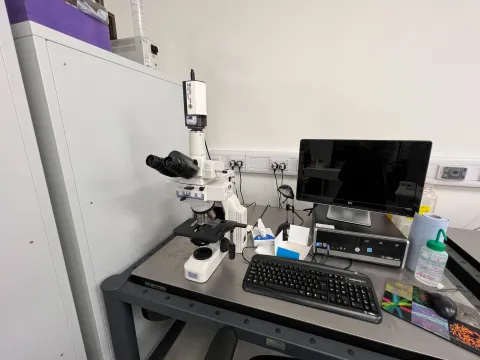
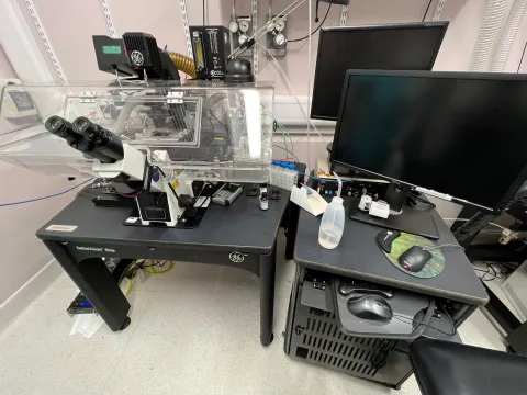
About the Confocal microscopes

天发娱乐棋牌_天发娱乐APP-官网|下载 confocal microscopes provide high magnification and high resolution imaging.

The principle difference between confocal microscopy and conventional widefield fluorescence microscopy is that images are comprised of stacks of sections of a discrete thickness. These can be used for 3D microscopy.

Sections of a known thickness are also superior for making quantitative measurements such as protein expression. Confocal microscopes are also slightly higher resolution than conventional widefield microscopes, and provide a sharper, higher SNR image.

Part of: Imaging and microscopy centre

Technical specification

Leica STELLARIS Confocal microscope

  • white light laser providing a continuous choice of excitation wavelengths  into NIR (485-790 nm)
  • 405 nm CW laser
  • 448 nm CW laser
  • 5 x “Power HyD” GaAsp detectors, allowing up to 5 channel simultaneous imaging when used with additional NIR capability
  • “super-resolution” confocal to 120 nm resolution.
     
Objectives:
  • 10x, 0.4, N.A.
  • 20x, 0.75, N.A.
  • 40x, 1.25, N.A., Glycerol with motorised correction collar
  • 63x, 1.4, N.A., Oil


“TauSense” uses fluorescence lifetime detection for additional fluorophore separation, or distinct gating of overlapping signals into separate channels dependent on their lifetime, for example autofluorescence.

Changes in a fluorophores nano-environment can also be measured using fluorescence lifetime e.g. FRET of interacting molecules, pH etc.


Features
  • transmitted light
  • DIC
  • hardware autofocus
  • simultaneous fluorescence acquisition
  • sequential fluorescence acquisition 
  • mark and find
  • motorised stage with Tile Scan (stitch multiple FOV together)
  • time-lapse
  • photo-kinetics
  • FLIM
  • NIR Imaging
  • lambda-scan/spectral channel un-mixing
     

2 x Leica SP8 AOBS Laser Scanning Confocal Microscopes

Modality and resolution
  • CLSM - 200 nm
  • DIC - 250 nm
Objectives
Magnification, numerical aperture and immersion
  • 10x, 0.5, air
  • 20x, 0.7, air
  • 40x, 1.3, oil
  • 63x, 1.3, glycerol
Specimen format
  • standard glass slide
  • 35 mm coverslip bottom dish
  • coverslip bottom chamber-slide
  • optical bottom multi-well plate
Detectors
  • confocal 1: 2 x PMT (photomultiplier tube), 1 x “HyD” GaAsP detector
  • confocal 2: 1 x PMT, 2 x “HyD” GaAsP detector
Lasers/filters
Excitation laser λ
  • 405 nm  (nano meters)  
  • 458 nm    
  • 476 nm    
  • 488 nm    
  • 496 nm    
  • 514 nm    
  • 561 nm    
  • 594 nm    
  • 633 nm   
Emission filters
  • 3 x Spectral Detectors. Any λ/Bandwidth from 400-800 nm. 
Fluorescence acquisition
  • simultaneous fluorescence acquisition of up to 3 channels
  • sequential fluorescence acquisition is unlimited
Other features
  • transmitted light
  • differential interference contrast (DIC)
  • time lapse
  • photo-kinetics
  • 3D imaging

3D specimen penetration is less than 100 μm.

The microscope does not have hardware autofocus. It has environmental temperature and gas control and a motorised stage with tile scan (stitch multiple FOV together).

It also offers mark and find and lambda-scan and spectral channel un-mixing.

Contact us

Dr Mark Willett

Highfield

Building 85, Highfield Campus, 天发娱乐棋牌_天发娱乐APP-官网|下载 SO17 1BJ
We’re open Monday to Friday 09:00 to 17:00 UK time.